Vascular IVR
Products
-
Preoperative simulation
Vascular model
Geometry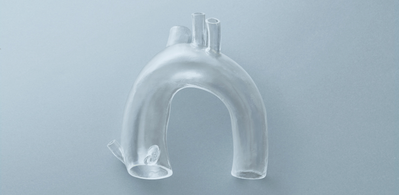
We provide transparent and accurate vascular models at a reasonable price.
More details


-
Preoperative simulation
Vascular Model
Custom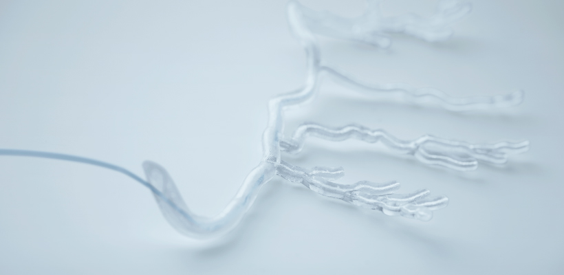
We provide transparent and highly accurate blood vessel models based on DICOM data.
For more information, click here.
-
Medical simulator
Repair and customization
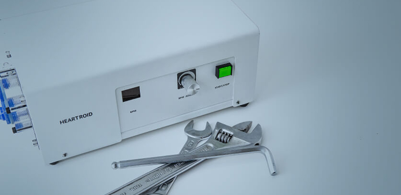
The simulator can be analyzed and repaired with a high-precision industrial CT scan.
More details


-
Preoperative simulation
HEARTROID
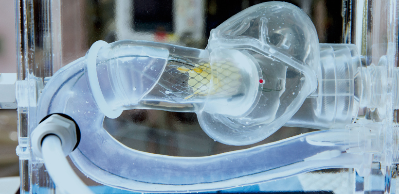
Cardiac Catheter Training System
More details


-
Preoperative simulation
Neurovascular
model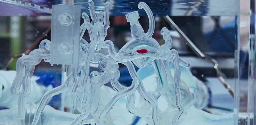
Cardiac Catheter Training System
More details
-
Preoperative simulation
HEARTROID
Peripheral Series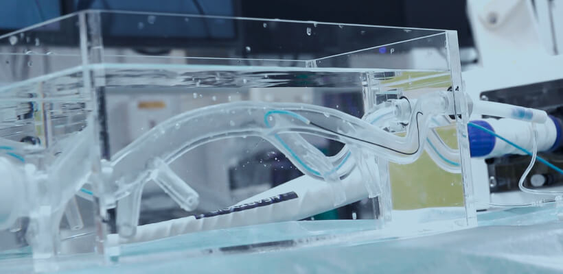
Cardiac Catheter Training System
More details
-
Preoperative simulation
Multi-functional pulsating pump
EC Series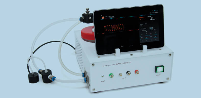
This is a pump system that can easily generate steady and pulsating flow.
More details


-
Preoperative simulation
4ch pressure display unit
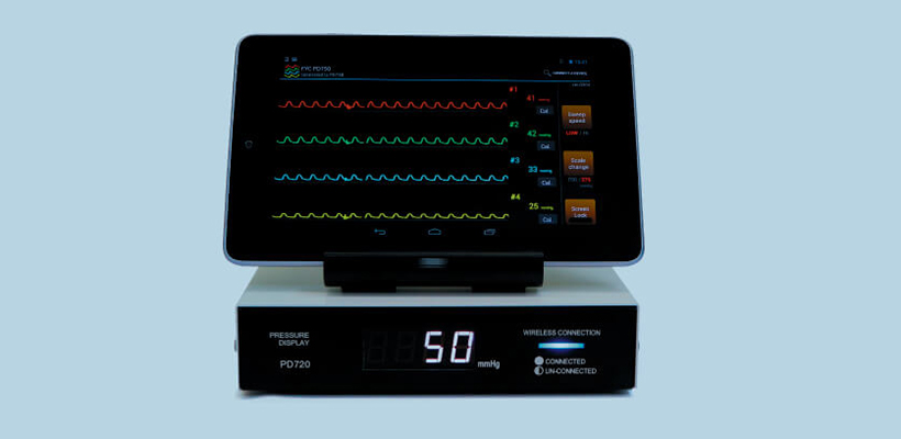
Pressure at up to four locations can be displayed simultaneously.
More details


From CT imaging to 3D printer output
- Reads DICOM data obtained from CT imaging into mimics
- Eliminates noise (artifacts) generated by metal and other materials
-
3D data is converted to images or data to see if the region of interest is
We will confirm with the customer that the area of interest has been reproduced.
The customer will be asked to confirm that the area of interest is reproduced in the image or data.If the STL output is available directly from the medical workstation, the above process is eliminated.
- 3D printing


Use of 3D Printers
近年は歯科用CTや医療用CTの進化と普及により、患者さまご自身のデータ(歯列と骨格)を取得することが出来るようになりました。CT撮影で得られたデータを加工し3Dプリンターで出力することにより術前のシミュレーションを実物大モデルでおこなえるようになりました。実物大モデルを手にすることでCAD画面上では不可能なフィット感などを治療チームで共有することにより、高度な治療計画を練ることができます。

