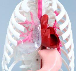
Bronchial preoperative simulation model
The bronchial simulator can be used as an educational tool for students and young doctors to understand
the anatomy of the bronchi. The bronchoscopy simulator can also be used for examination and treatment
devices.
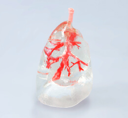
This model is manufactured using a 3D printer, so it can reproduce up to 23 branches, the
maximum number of branches.
Reproducible range
The bronchial model faithfully reproduces the bifurcation of the bronchi, allowing for both
bronchoscopic examination and the use of therapeutic devices.
Possible range of reproduction: Air duct, upper lobe air duct branch, middle lobe air duct branch,
lower lobe air duct branch
Reproducible branches: 23 branches (peripheral)
Advantages of using a bronchial simulator
Main uses
We envision validation models for clinical settings, medical device operation practices, and medical
device development.
- Educational model for understanding anatomy for students and young doctors
- Bronchoscope operation practice
- Training on treatment devices
- Functional evaluation at the development stage of medical devices
Supported Materials
The material can be selected up to the third branch of the bronchus, but after that, the tip of the
bronchus is made of hard material.
Contact Us