Tooth transplant simulation using optical modeling
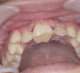
In the past, tooth transplant surgeries required the use of x-rays and CT to confirm the shape of the
tooth to be transplanted, such as wisdom teeth, and to shave the jawbone and tooth to be transplanted at
the time of surgery. At this time, if the transplanted tooth is repeatedly removed and inserted into the
site of the transplant, the area called the periodontal ligament is damaged, and the rate of retention
after transplantation tended to be low.
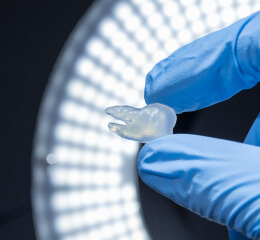
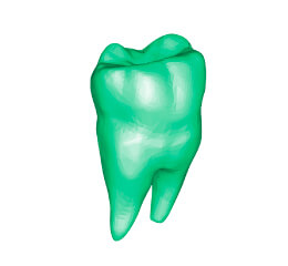
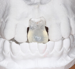
Wisdom teeth to be used as transplanted teeth are photographed with a CT scanner, reconstructed as 3D
data, and fabricated into an elaborate three-dimensional model using optical modeling. CT and MRI images
can be converted into 3D data, which can then be fabricated into a three-dimensional model using optical
modeling.
In implant treatment
Advantages of using 3D printers
There are four major advantages to confirming the depth and angle of implant placement in advance.
-
Shorter treatment time
-
Improvement of treatment accuracy
-
Reduce the burden
on the patient
-
Improvement of techniques
Main uses
- Preoperative simulation
- Confirmation of procedures and treatment plans
- Training of trainees and students
Contact Us