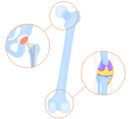
Femur Fracture Treatment Simulation Model
The femur is located in the thigh and is one of the most important bones for walking. Fractures of the
femur include fractures of the head, neck, and posterior portion of the femur, of which fractures of the
head and neck at the hip joint require special attention.
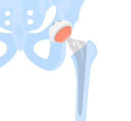
The hip joint area is where the elderly often develop osteoporosis and fragility fractures, which can go
unnoticed and cause muscle weakness and risk of complications such as pneumonia as people stop walking
to avoid pain.
In addition, the arteries in the bone head and neck are thin, and if damaged, they can cause
"osteonecrosis of the femoral head," in which bone cells die, or "delayed ossification," in which the
bone head collapses. These require surgery as early as possible, and procedures such as
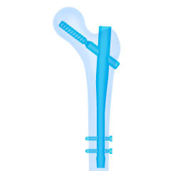
"osteosynthesis," in which the patient's own bone is transplanted, and "artificial head insertion," in
which the bone is replaced with an artificial one, are performed.
JMC Lab's preoperative simulator can produce a full-scale model of the patient and verify the position
of the graft before surgery.
Advantages of Femur Fracture Treatment Simulation Model
-
Shorter treatment time
-
Improvement of treatment accuracy
-
Reducing the burden
on patients
-
Improvement of techniques
Main uses
- Confirmation of procedures and treatment plans
- Training of trainees and students
- Development tools, sales tools, and marketing tools for medical device
manufacturers
Contact Us