3D Pringting
JMC Technology
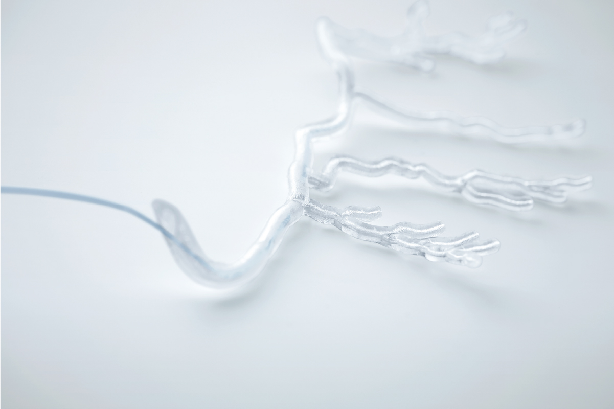
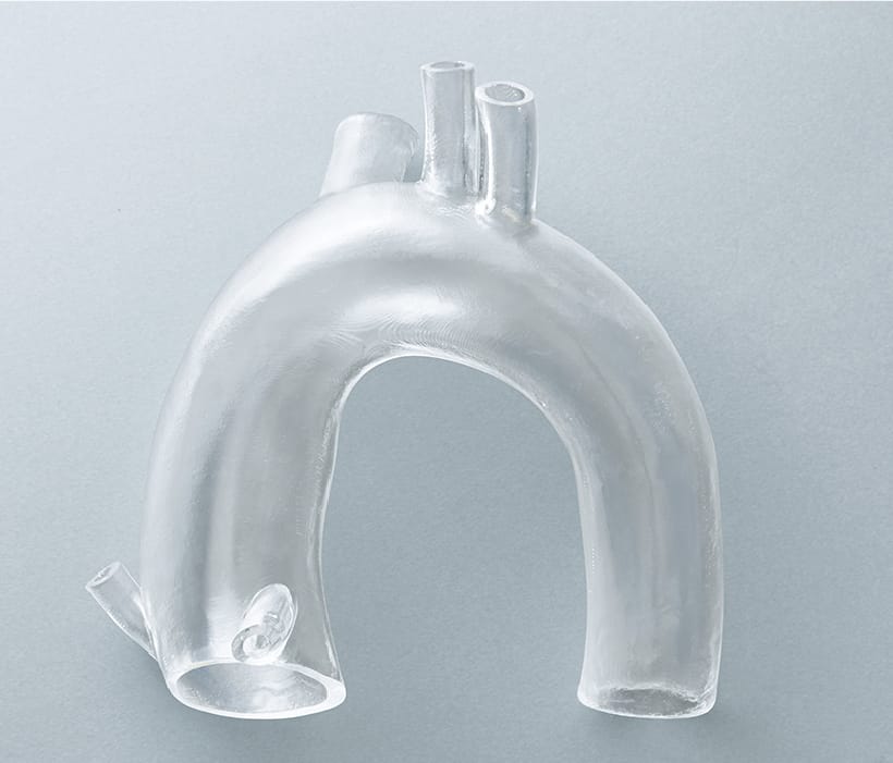
Soft model with unique technology
In general, soft models often fail to reproduce some shapes, such as simplifying the inside of organs, but JMC Lab possesses a unique technology that combines 3D printing and casting technologies to faithfully reproduce the outside and inside of organs. JMC Lab has a unique technology that combines 3D printing and casting techniques to faithfully reproduce the outside and inside of organs. This method allows for a higher degree of material flexibility than direct 3D printing of soft materials, and allows for adjustments in hardness, color, X-ray transmission, etc.
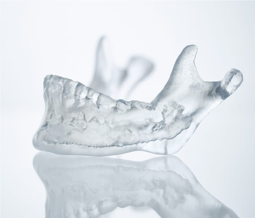
Rigid model with short delivery time
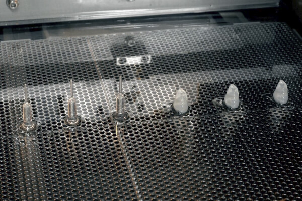
We have a system in place that allows us to work 24 hours a day, 7 days a week on rigid models that are output directly from the 3D printer, so that we can deliver them quickly to our customers in fields that require speed, such as preoperative simulation and prototyping of medical devices. We have a variety of output methods, including optical, SLS, and inkjet, so you can choose the most suitable method for your application.
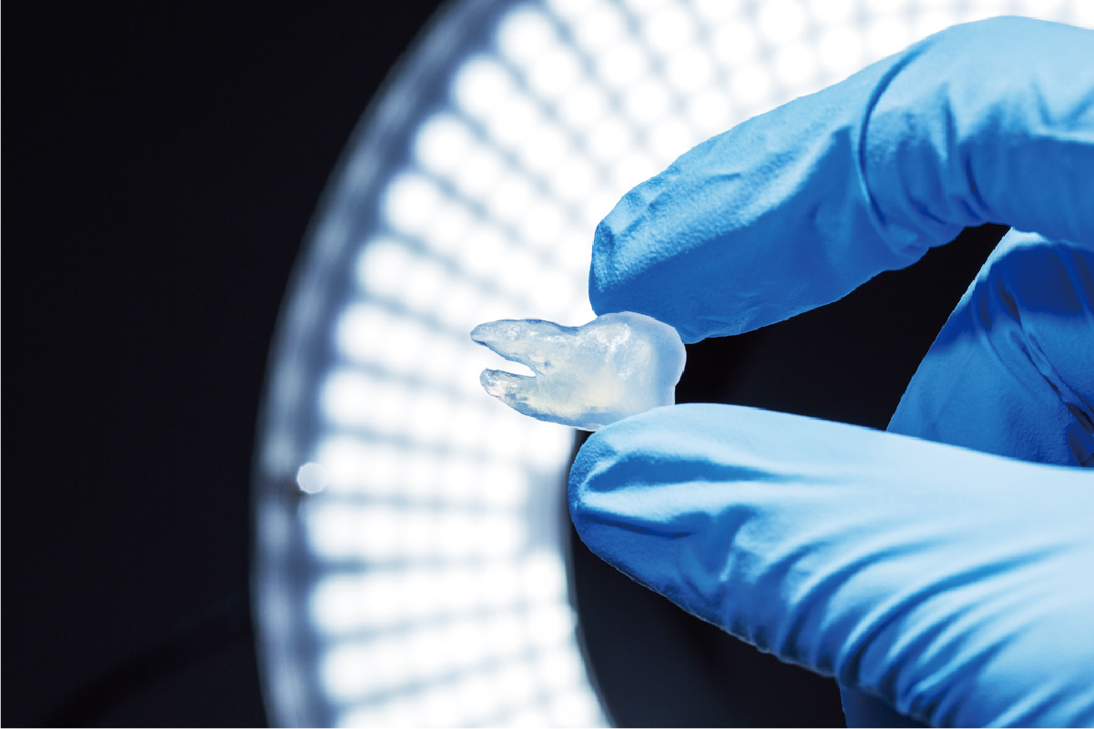
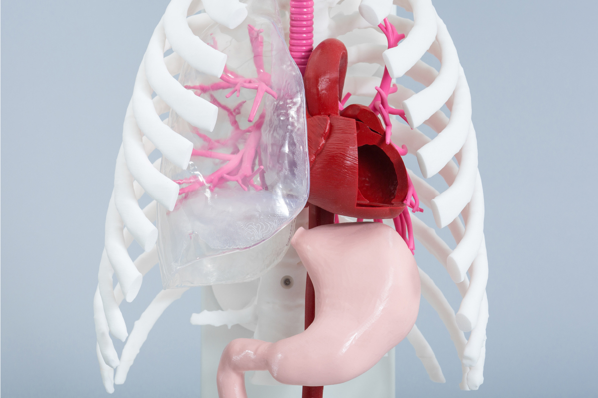
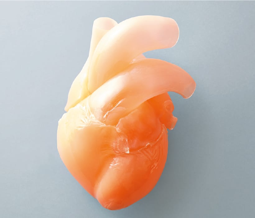
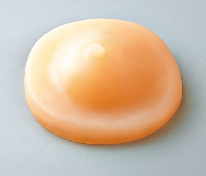
Soft organ model
One of the strengths of JMC's medical entity models is the ability to produce soft models. However, JMC's unique know-how and painstaking manufacturing methods enable us to accurately reproduce the complex shapes of organs and to adjust the hardness according to the part to be manufactured. JMC's unique know-how and labor-intensive manufacturing methods enable us to accurately reproduce the complex shapes of organs and adjust the hardness of the parts we manufacture.
All models are customizable.
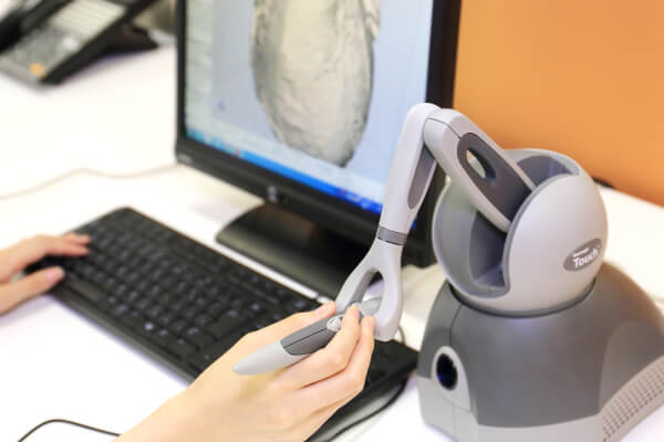
JMC all of our models are made to order. In addition to directly sculpting the customer's 3D data, we can also customize the data and create data from CT and MRI. Even if you do not have any original data, we can make a proposal to realize your image based on your sketch.
Data editing
Model Surgery
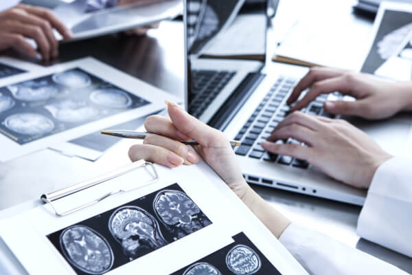
- Reads DICOM data obtained from CT imaging into mimics
- Eliminates noise generated by metal and other materials
-
3D data is converted to images or data to see if the region of interest is
We will confirm with the customer that the area of interest has been reproduced.
The customer will be asked to confirm that the area of interest is reproduced in the image or data.If the STL output is available directly from the medical workstation, the above process is eliminated.
- 3D printing
About DICOM data
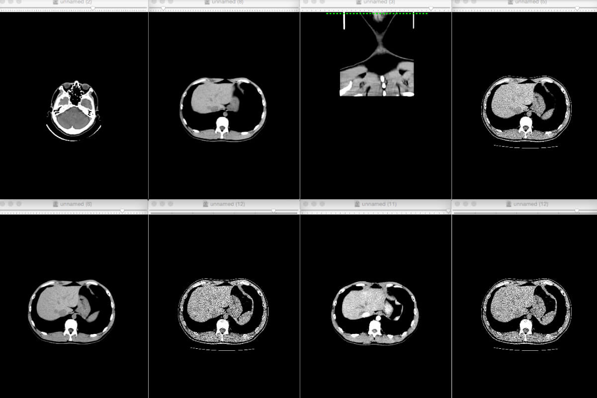


3D data extraction from medical CT and MRI data
3D data is essential for the production of models using 3D printers, but it is not always possible to
prepare 3D data in the medical field. In such a case, if you can provide us with DICOM data obtained from
CT or MRI, we can create precise 3D data.
JMC uses a software called "mimics" to construct precise skeletal and organ shapes from DICOM data. JMC
uses mimics software to construct precise shapes of skeletons and organs from DICOM data. By having
specialized engineers perform image processing using mimics, it is possible to extract only the areas of
interest and output higher quality 3D data with less distortion caused by metal products such as gold
teeth and pacemakers.
Supported materials
-
rapid prototyping (machine)
Complex shapes can be integrally molded in a short time. Since the liquid resin is cured by the laser one layer at a time, the "corner radius" (radius of a cylindrical tool) that occurs in blade processing is not generated.
This makes it possible to reproduce the microscopic shape of organs.
-
Silicone
Materials from a variety of domestic and overseas manufacturers can be used. In addition to the characteristics of high transparency and low hardness, it is possible to adjust the hardness by using silicone thinner.
For some silicones, it is possible to add more than 50 wt% thinner, but this will significantly reduce the physical properties, resulting in poor curing or thinner components bleeding onto the surface.
-
Polyvinyl alcohol(PVA)
The material itself, called hydrogel, contains water, so it has good wettability and slipperiness, and feels very close to the organs of the human body.
-
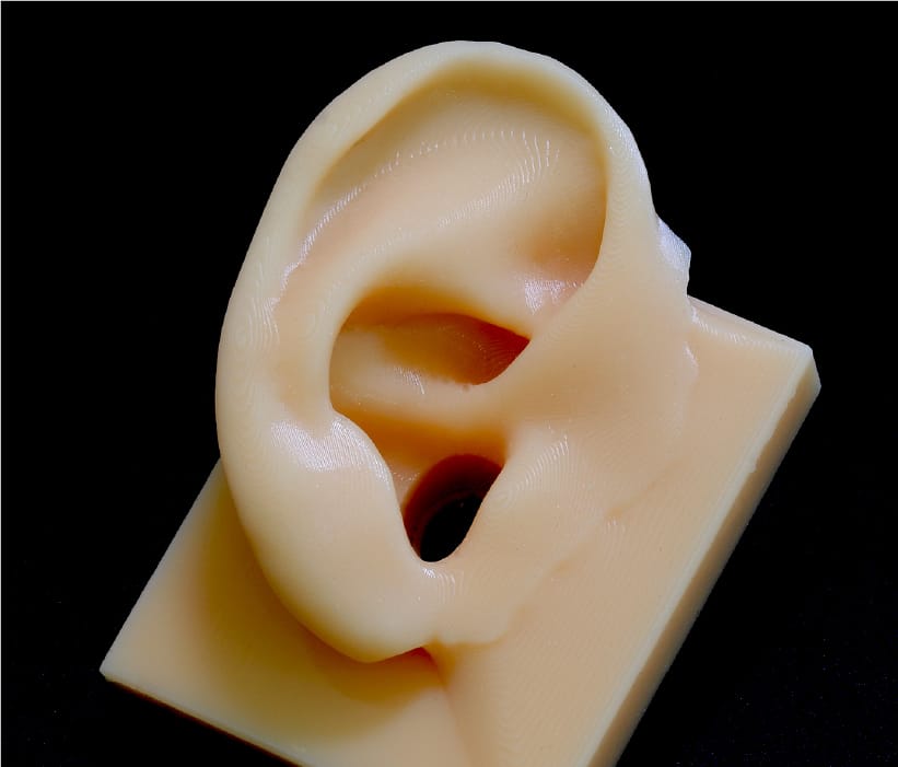
Urethane foam
It can reproduce both low and high hardness, which is similar to the touch of human skin.
-
Ultrasonic compatible materials
Materials that can be captured on an ultrasound imaging system can be used.
-
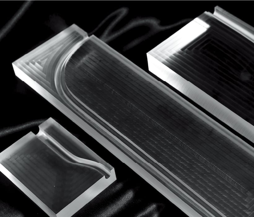
Cutting materials
Materials such as acrylic, ABS, polycarbonate, and polypropylene can be used.